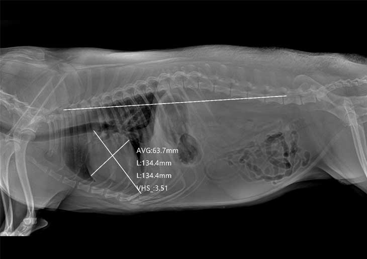
Veterinary DR examination is an important diagnostic method that uses DR X-ray equipment to observe the anatomical morphology, physiological function, and pathological changes of internal organs in the body, thus diagnosing various diseases. This method includes fluoroscopy, radiography, and contrast examinations.
Veterinary Fluoroscopy Examination

Connect the animal DR and its accessories as per the instructions in the manual.
Adjust the machine settings. Typically, use a tube current of 2-3 milliamperes, and the tube voltage should be determined based on the thickness of the area being examined, with 50-70 kilovolts being suitable. The distance between the machine and the animal is generally set between 50-100 centimeters.
Clean the animal's body, remove any items on them, and secure them properly. Administer sedatives or general anesthesia as needed.
Wear protective gear.
Position the fluorescent screen close to the animal being examined. Change the animal's position if necessary to get a comprehensive view of the area of interest.
Turn on the machine, use intermittent exposure, and make use of collimation to minimize the examination time.
Understand the basic information of the dog or cat, as well as the imaging requirements and purposes.
Clean the animal's body, remove any items, secure them properly, and administer sedatives or general anesthesia if necessary.
Select appropriately sized films based on the imaging area, measure the thickness of the area to be imaged, and determine imaging conditions such as tube voltage, tube current, exposure time, and distance.
Position the darkroom so that the center of the X-ray beam, the center of the area being examined, and the center of the darkroom are in a straight line.
Turn on the machine and expose while the dog or cat being examined is calm to obtain latent images.
Obtain digitized X-ray images.
Contrast imaging techniques are mainly used for tissues and organs in the digestive tract, urinary tract, bronchial tubes, and other areas where natural contrast is lacking.
Animal DR Digestive Tract Contrast Imaging:
Prior to the contrast imaging of the dog or cat, fast them and withhold water for at least 12 hours.
Mix barium sulfate with Arabic gum and add a small amount of hot water, followed by an appropriate amount of warm water. Use 60% barium sulfate for esophageal contrast and 15% barium sulfate for gastrointestinal contrast. Administer the barium contrast orally, with a dosage of 2-5 milliliters per kilogram of body weight.
Depending on the situation, you can choose to perform the examination in standing lateral position, dorsal lateral position, dorsal ventral position, or supine position. The examination of the esophagus and stomach can be observed immediately or shortly after contrast administration, while the examination of the small intestine should be observed 1-2 hours after the barium administration, and the examination of the large intestine should be observed 6-12 hours after barium administration.
For bladder contrast imaging, insert a urinary catheter, empty the urine, and then inject sterile air or 10%-20% sodium iodide into the bladder, with a dose of 6-12 milliliters per kilogram of body weight.
For renal pelvis contrast imaging, the dog or cat should fast for 24 hours and refrain from drinking water for 12 hours before the examination. They should be in a supine position with a pressure band placed on the lower abdomen to prevent the contrast agent from entering the bladder and affecting renal pelvis filling. Slowly inject 50% contrast medium or 58% urografin, at a dosage of 20-30 milliliters, via intravenous injection. After the injection, take abdominal and dorsal images 7-15 minutes later. Once the renal pelvis is imaged, remove the pressure band and then take images of the bladder.
Bronchial Tube Contrast Imaging:
The dog or cat being examined should be placed in a lateral recumbent position.
Insert a catheter through the mouth or inject the contrast agent directly into the trachea (approximately 15 milliliters of 40% iodinated poppyseed oil). The contrast medium flows into the bronchial tube along the lower bronchus. Under fluoroscopy, you can observe the contrast medium flowing sequentially into the lobular bronchi, diaphragmatic bronchi, and apical bronchi. Once the contrast medium has completely entered the bronchial tube, take X-ray images.
Only one side of the lung bronchi can be examined at a time. If a contrast imaging of the opposite bronchus is needed, it should be performed after the contrast medium has been completely expelled.