T9-VET is a high-end veterinary color doppler ultrasound tailored for pet hospitals, scientific research institutions, university and college, animal research institutes, pharmaceutical companies, etc Meet the cat, dog, mouse, rabbit, and other small pets' cardic, pregnancy, digestive system, reproductive system, urinary system, cardiovascular and other aspects of the examination and diagnosis.
Excellent performance, excellent reliability
Powerful function to meet the needs of inspection and diagnosis. Extremely strong penetration, uniform and detailed image. Integrate the sense of science and technology, humanized design concept, senior introverted,and intelligence and beauty all in one. Multi-channel interoperability full activation probe interface, built-in data storage space and other hardware configurations.
ST-K advanced algorithm
COM-E high-end hardware platfor
CPU+GPU high-speed computing power
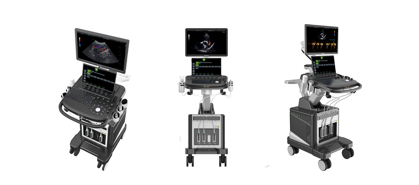
Appearance Display
High-end design
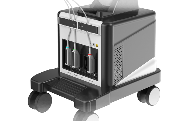
Four-probe interface
Single crystal technique
senior introverted ,wisdom & beauty are one
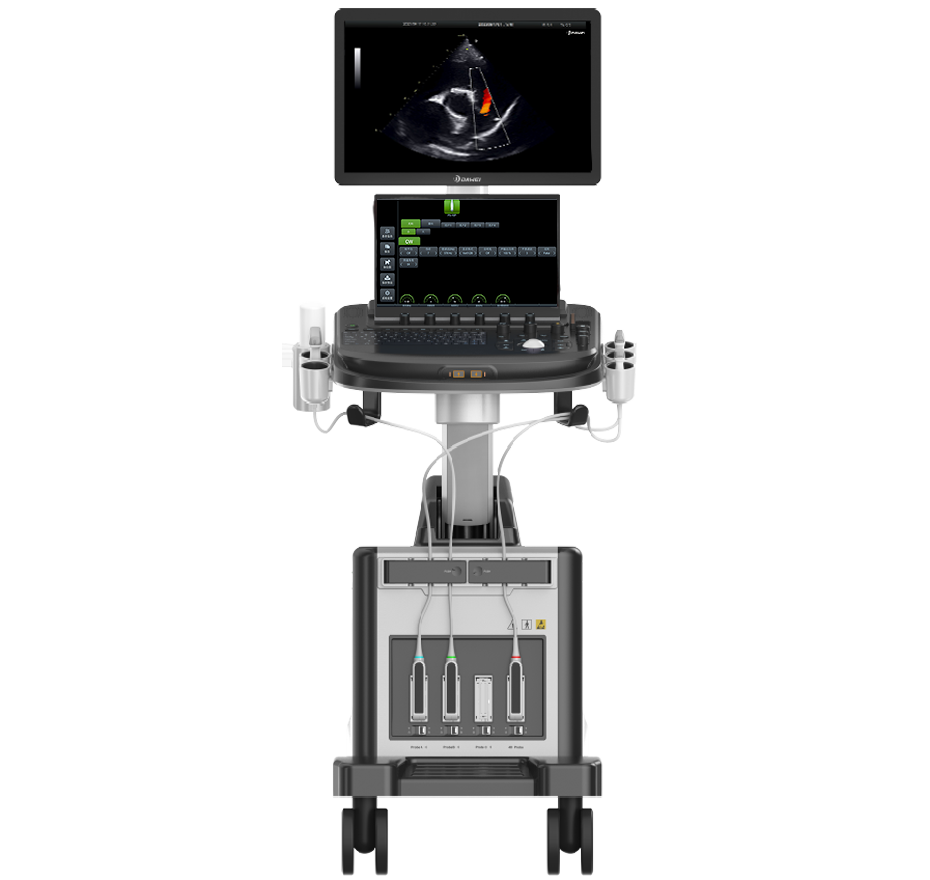
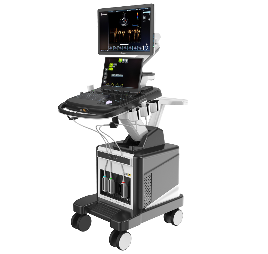
110° Left-right Angle Adjustment Electric Height Adjustment
21.5-inch Medical High-definition Display
15.6-inch Extra-large Touchscreen
Fully activated four-probe interface
Meet the clinical application requirements of various departments
ST-U Single Crystal Probe
High-End Single Crystal Technology, Delivering Higher Energy Conversion Efficiency
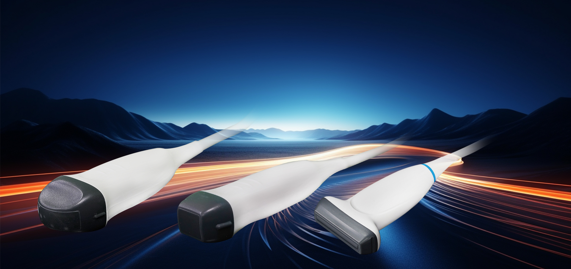
Probe
Advanced Probe Material Applications
The probe is made from composite materials, offering superior penetration and higher resolution. The images are consistently uniform without any loss of detail, providing more than a 60% improvement in performance compared to traditional piezoelectric ceramic materials.
Sub-cutting allows for complete control of the entire process of chip vibration, reducing sidelobe artifacts, enhancing fine tissue resolution, and achieving sharper boundaries between adjacent strong echo reflectors. This fully showcases the high-resolution images brought by the high-density probe, perfectly presenting image details, and increasing clinical diagnostic accuracy.
Lossless Signal Transmission and Reception
High Precision Mode Switching
Spatiotemporal Wave Velocity Reconstruction
Image Post-Processing
Unique pure wave probe technology
Bring high resolution images
Featuring a dedicated high-density probe, employing a completely new array design technology and an unique pure wave probe technology, undergoes secondary cutting of individual transducers, allowing for full control over the transducer's vibration throughout the process. This reduces sidelobe artifacts, enhances the resolution of fine tissue details, and sharpens the boundaries between adjacent strong echo reflectors. It fully showcases the high-resolution images brought by the high-density probe, perfectly presenting image details, and increasing the accuracy of clinical diagnosis.
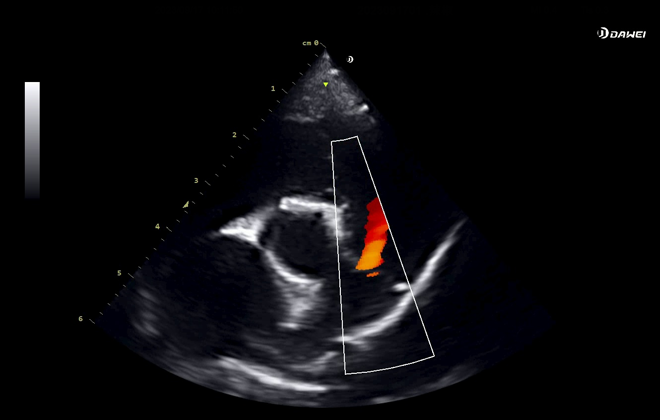
Clinical Images
maging Technology
Pulmonary Artery Regurgitant Flame Sign
Micro-convex Longitudinal View of Liver Vasculature
Renal Blood Flow with Micro-convex Effect
Linear Array Left Ovarian Effect
Right Lateral Decubitus Mitral Valve Short-axis M-mode Effect