Veterinary Color Doppler Ultrasound Diagnostic System
latest technology chip design for a wide range of applications
Veterinary Clinic
Animal Research Institute
Livestock Husbandry College
Product Advantages
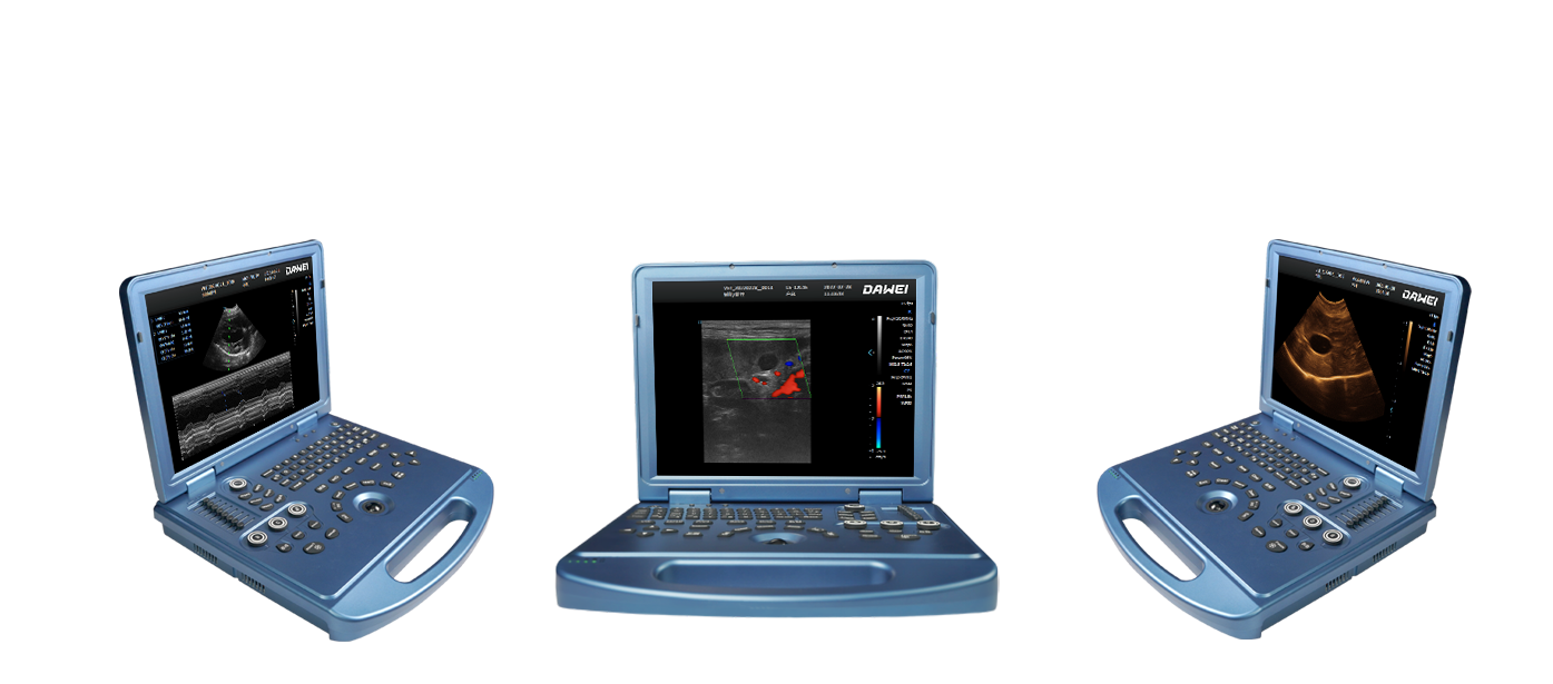
L5-VET veterinary color doppler system with highly integrated hardware modules, lightweight and high image effects, very portable for outdoor diagnosis. Small external size built-in large capacity lithium battery, providing effective protection for outdoor diagnosis. Smooth lines design of the unit, exquisite and attractive appearance
Unique YH platform
Adopting a variety of advanced chip technologies with highly integrated hardware modules, the YH platform greatly improves system's computing power and processing capacity, making ultrasound diagnostic operations more convenient and efficient, and providing more confidence for clinical diagnosticians。
Harmonic Imaging Technique
High Definition Liver Imaging Effects
Trapezoidal Imaging Technique
Appearance Display
Appearance
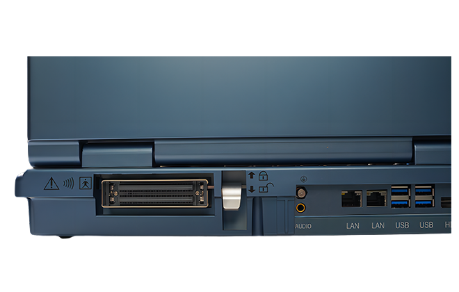
Extensive External Interfaces
Technical Features
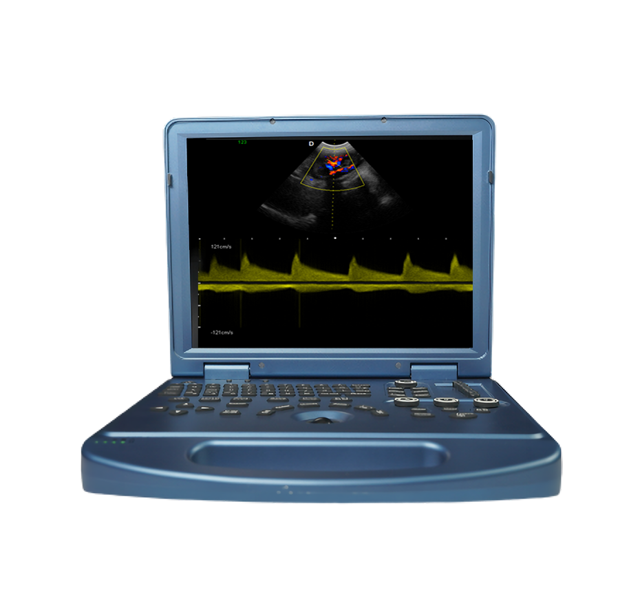
Intelligent and efficient lightweight and portable
15-inch Medical High-Definition Display
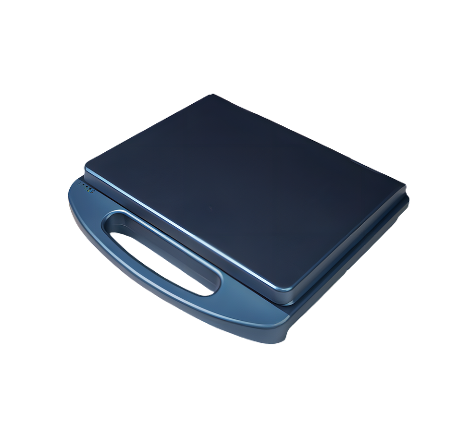
Slim and Lightweight Body
Abundant External Interfaces
Plug to use
5*USB ports、1*Audio、1*HDMI、2*RJ-45
Trapezoidal Imaging
Trapezoidal imaging is kind of extended imaging, on the basis of the original rectangle is converted into a trapezoidal shape, the left and right sides of a certain degree of expansion, to achieve a wider field of view effect。
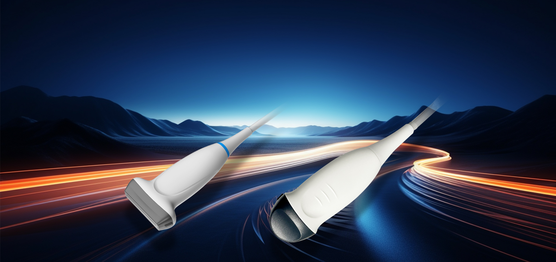
Probe
Advanced Probe Applications
The probe is made from composite materials, offering superior penetration and higher resolution. The images are consistently uniform without any loss of detail, providing more than 60% improvement in performance compared to traditional piezoelectric ceramic materials。
Sub-cutting allows for complete control of the entire process of chip vibration, reducing sidelobe artifacts, enhancing fine tissue resolution, and achieving sharper boundaries between adjacent strong echo reflectors. This fully showcases the high-resolution images brought by the high-density probe, perfectly presenting image details, and increasing clinical diagnostic accuracy.
Lossless Signal Transmission and Reception
High Precision A/D Conversion
Spatiotemporal Wave Velocity Reconstruction
Image Post-Processing
Unique pure wave probe technology
Bring high resolution images
Featuring a dedicated high-density probe, employing a completely new array design technology and an unique pure wave probe technology, undergoes secondary cutting of individual transducers, allowing for full control over the transducer's vibration throughout the process. This reduces sidelobe artifacts, enhances the resolution of fine tissue details, and sharpens the boundaries between adjacent strong echo reflectors. It fully showcases the high-resolution images brought by the high-density probe, perfectly presenting image details, and increasing the accuracy of clinical diagnosis.
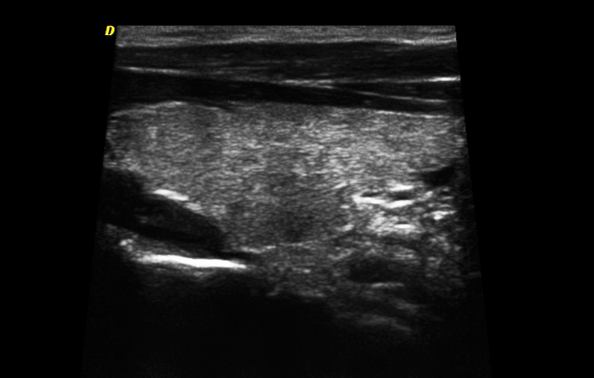
Clinical Images
Imaging Technology
Liver and Gallbladder Pseudo-color Micro-convex Section