Veterinary Color Doppler Ultrasound Diagnostic System
Variety of imaging functions for meeting the diagnostic needs of various scenarios
Animal research institute
Veterinary Clinic
Breeding&propagation base
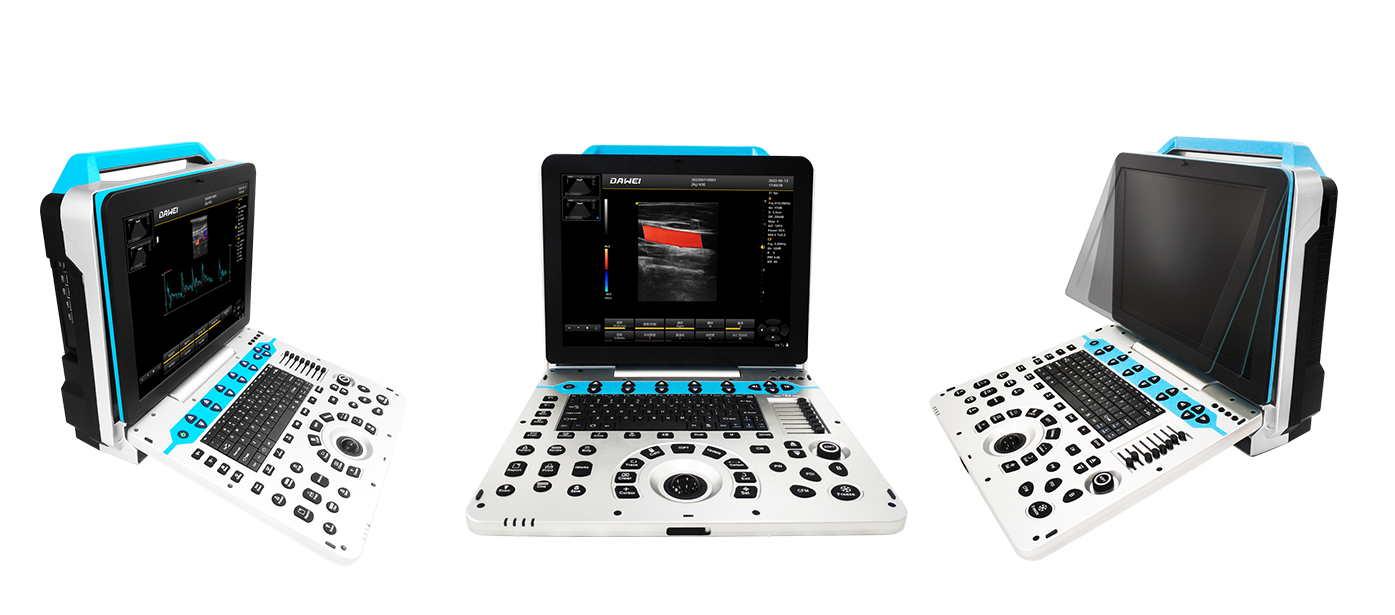
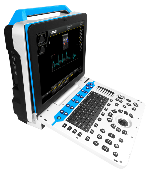
Appearance Display
Appearance
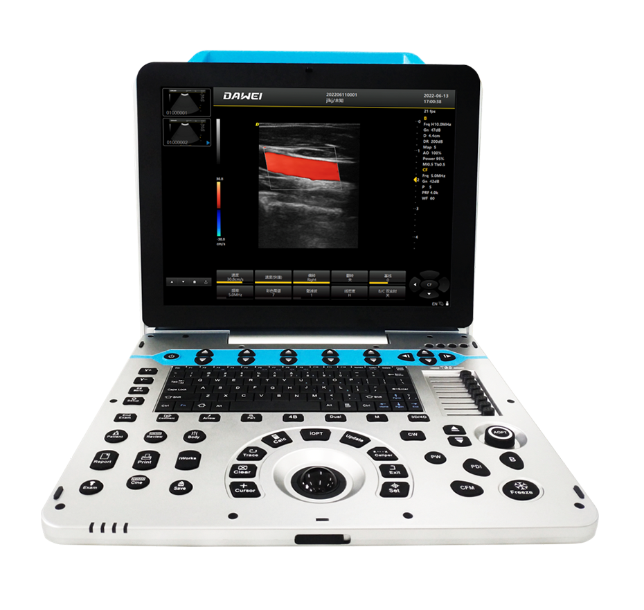
Ultra-high Definition Large Screen
Technical Features
Satisfy the Meed for A User-friendly Control Experience
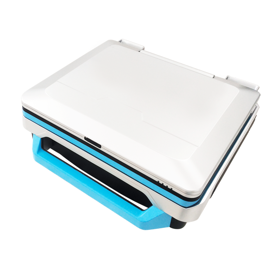
High-Capacity Removable Lithium Battery
0-30° Adjustable Display Angle
Fully Activated Dual Probe Interface
15-inch High-Resolution Display
Spatial Composite Imaging Technology
Ultrasound Spatial Compound Imaging can enhance contrast resolution, fine resolution, and spatial resolution; improve tissue and lesion interface echo continuity, and reduce various artifacts (mirror reflection, speckle, scattering, attenuation, and contrast differences)
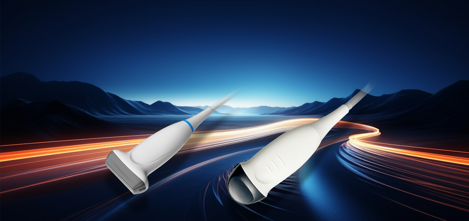
Probe
Advanced Probe Applications
The probe is made from composite materials, offering superior penetration and higher resolution. The images are consistently uniform without any loss of detail, providing more than 60% improvement in performance compared to traditional piezoelectric ceramic materials。
Sub-cutting allows for complete control of the entire process of chip vibration, reducing sidelobe artifacts, enhancing fine tissue resolution, and achieving sharper boundaries between adjacent strong echo reflectors. This fully showcases the high-resolution images brought by the high-density probe, perfectly presenting image details, and increasing clinical diagnostic accuracy.
Lossless Signal Transmission and Reception
High Precision A/D Conversion
Spatiotemporal Wave Velocity Reconstruction
Image Post-Processing
Unique pure wave probe technology
Bring high resolution images
Featuring a dedicated high-density probe, employing a completely new array design technology and an unique pure wave probe technology, undergoes secondary cutting of individual transducers, allowing for full control over the transducer's vibration throughout the process. This reduces sidelobe artifacts, enhances the resolution of fine tissue details, and sharpens the boundaries between adjacent strong echo reflectors. It fully showcases the high-resolution images brought by the high-density probe, perfectly presenting image details, and increasing the accuracy of clinical diagnosis.
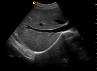
Clinical Images
Imaging Technology
Color Doppler Blood Flow Effect
Bladder Microconvex Section
Renal Micro-convex Section
Renal Linear Array Color Doppler Blood Flow Imaging
Short-axis Apical View of The Left Ventricle of The Heart