P60-VET veterinary color doppler system utilizes the innovative ST-U Rapid Imaging Platform for continuous high-definition images integrating advanced CPU+GPU high-speed algorithms with a tech-savvy and user-friendly design philosophy, upgrading imaging power through enhanced hardware configurations。
Rich technical functionalities
Outstanding mobility, capable of handling various complex environments, and can meet the needs of scientific research and teaching units, zoos, aquatic research institutes, veterinary clinics, breeding organizations, etc。
Speckle Reduce Imaging
Real-time dual image contrast display
Color hiding technology
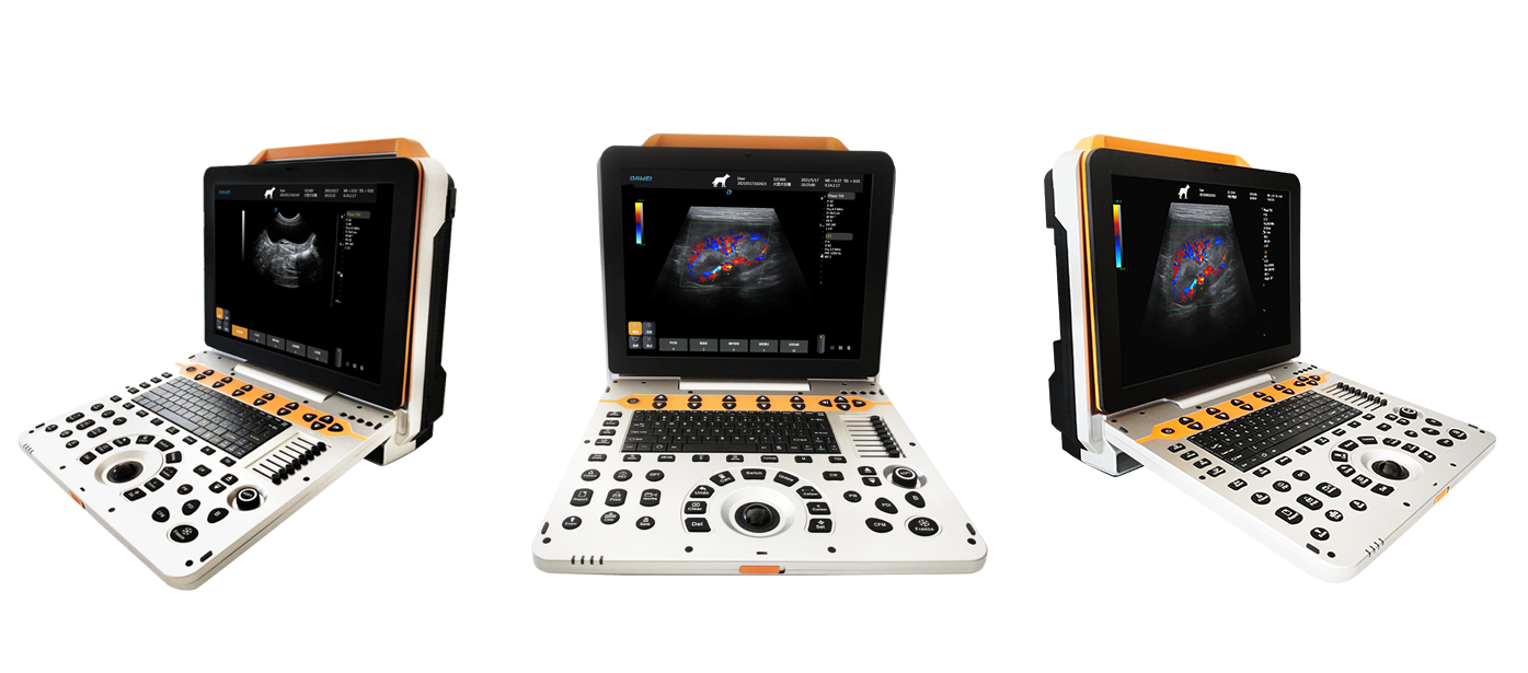
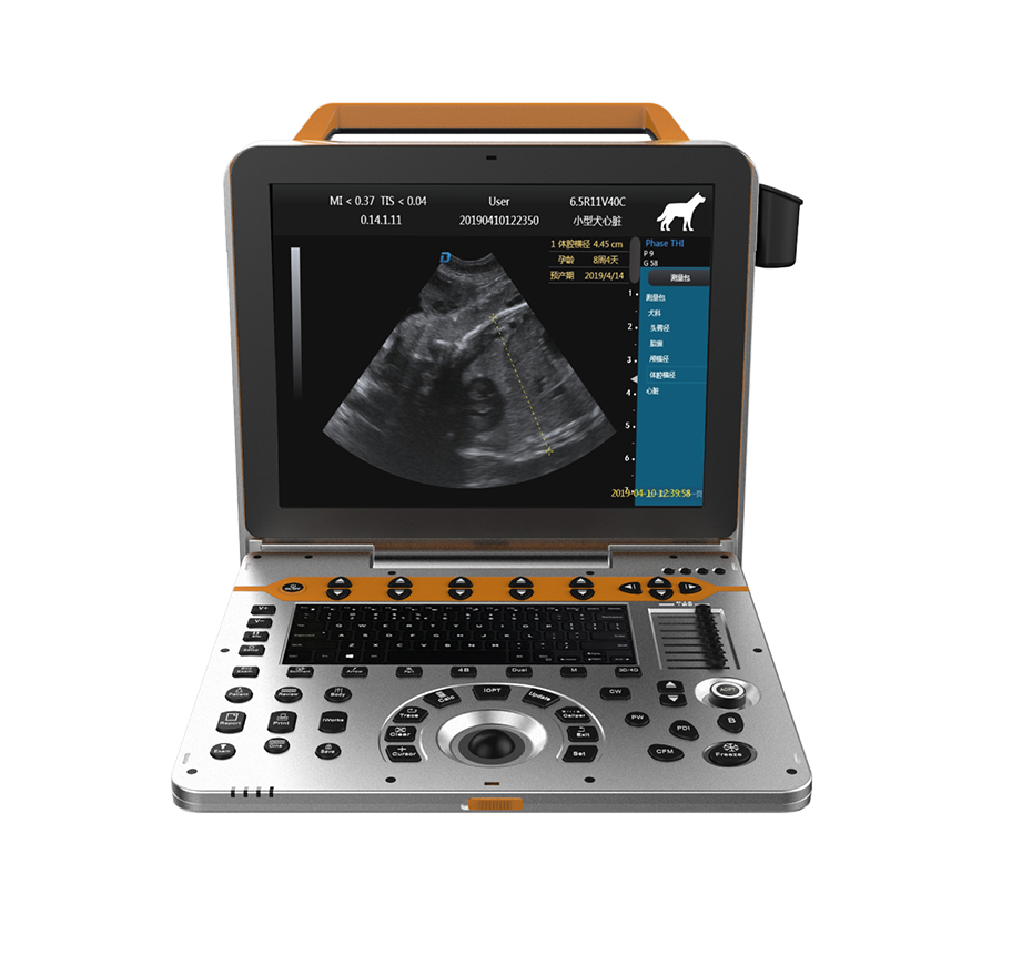
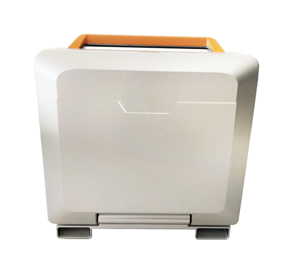
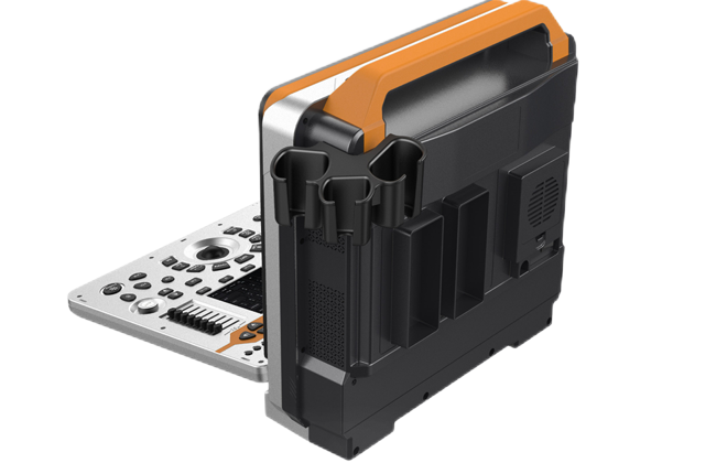
Appearance Display
Appearance
Multi-Transducer Interface
Technical Features
Deep Technology Empowering the Future Around the Clock
Flexible Adjustment Of Display Within 45°
Equipped With High-End Imaging Technology
Main Screen 15-Inch High-Resolution Color Lcd Display
22 Built-in Probe Interfaces, All Activated, Uniform in Size, Interchangeable, with Flexible Adjustment Within 45 Degrees for the Display Screen
PView Wide-Angle Imaging Technology
Wide-Angle Imaging involves capturing a series of two-dimensional cross-sectional images through the movement of a probe and then using computer reconstruction methods to stitch these two-dimensional images together into a continuous cross-sectional image.
Probe
Advanced Probe Applications
The probe is made from composite materials, offering superior penetration and higher resolution. The images are consistently uniform without any loss of detail, providing more than 60% improvement in performance compared to traditional piezoelectric ceramic materials。
Sub-cutting allows for complete control of the entire process of chip vibration, reducing sidelobe artifacts, enhancing fine tissue resolution, and achieving sharper boundaries between adjacent strong echo reflectors. This fully showcases the high-resolution images brought by the high-density probe, perfectly presenting image details, and increasing clinical diagnostic accuracy.
Lossless Signal Transmission and Reception
High Precision A/D Conversion
Spatiotemporal Wave Velocity Reconstruction
Image Post-Processing
Unique pure wave probe technology
Bring high resolution images
Featuring a dedicated high-density probe, employing a completely new array design technology and an unique pure wave probe technology, undergoes secondary cutting of individual transducers, allowing for full control over the transducer's vibration throughout the process. This reduces sidelobe artifacts, enhances the resolution of fine tissue details, and sharpens the boundaries between adjacent strong echo reflectors. It fully showcases the high-resolution images brought by the high-density probe, perfectly presenting image details, and increasing the accuracy of clinical diagnosis.
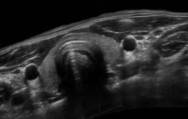
Clinical Images
Imaging Technology
Bladder Cross-sectional View
Adrenal PW Localization
Adrenal Abdominal Aorta PW Spectrum
Linear Array Trapezoidal Imaging of Renal Blood Flow
Linear Array Trapezoidal Imaging of The Pancreas Result