Veterinary Color Doppler Ultrasound Diagnostic System
Excellent performance software and hardware architecture
Compact Convenient
Intelligent control
Long endurance
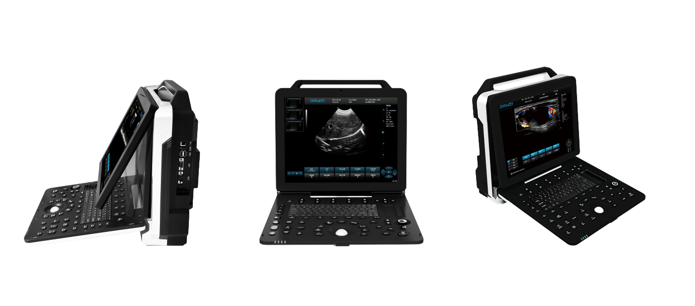
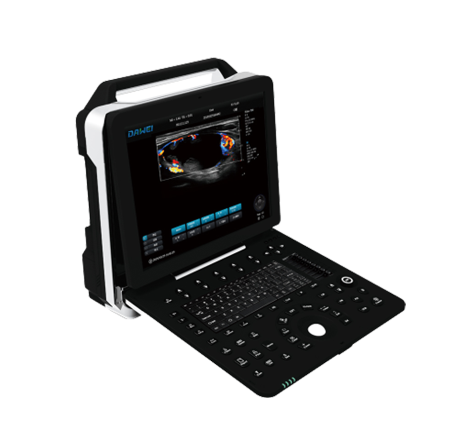
Appearance Display
Appearance structural design
Ultra-high definition large screen
Technical Features
More professional and comprehensive
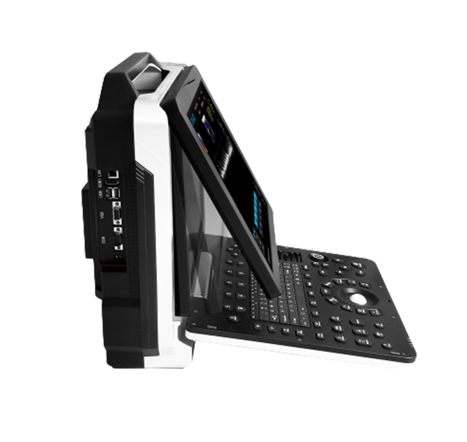
Flexible Adjustment Within 45 Degrees for the Display Screen
User-friendly Interactive Interface
Dual-Channel Probe Interface
Main screen is 15-inch medical HD display
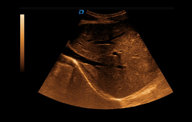
Contrast Tuned Imaging Technology
The intravenous contrast agent is injected into the body's blood circulation system, which has a strong acoustic impedance and enhances the contrast by the different way the sound waves reflect at the interface of different media, resulting in higher image resolution
Probe
Advanced Probe Applications
The probe is made from composite materials, offering superior penetration and higher resolution. The images are consistently uniform without any loss of detail, providing more than 60% improvement in performance compared to traditional piezoelectric ceramic materials.
Sub-cutting allows for complete control of the entire process of chip vibration, reducing sidelobe artifacts, enhancing fine tissue resolution, and achieving sharper boundaries between adjacent strong echo reflectors. This fully showcases the high-resolution images brought by the high-density probe, perfectly presenting image details, and increasing clinical diagnostic accuracy.
Lossless Signal Transmission and Reception
High Precision A/D Conversion
Spatiotemporal Wave Velocity Reconstruction
Image Post-processing
Unique pure wave probe technology
Bring high resolution images
Featuring a dedicated high-density probe, employing a completely new array design technology and an unique pure wave probe technology, undergoes secondary cutting of individual transducers, allowing for full control over the transducer's vibration throughout the process. This reduces sidelobe artifacts, enhances the resolution of fine tissue details, and sharpens the boundaries between adjacent strong echo reflectors. It fully showcases the high-resolution images brought by the high-density probe, perfectly presenting image details, and increasing the accuracy of clinical diagnosis.
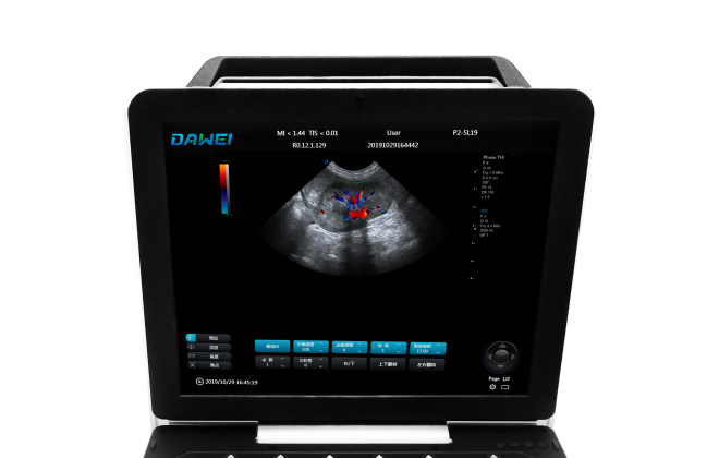
Clinical Images
Imaging technology
Color Doppler Hepatic Micro-convex Blood Flow Imaging
Color Doppler Renal Micro-convex Blood Flow Imaging