Veterinary Color Doppler Ultrasound Diagnostic System
Stylish Elegant Simple Extraordinary
Simple Stylish
New Architecture
Stable and Durable
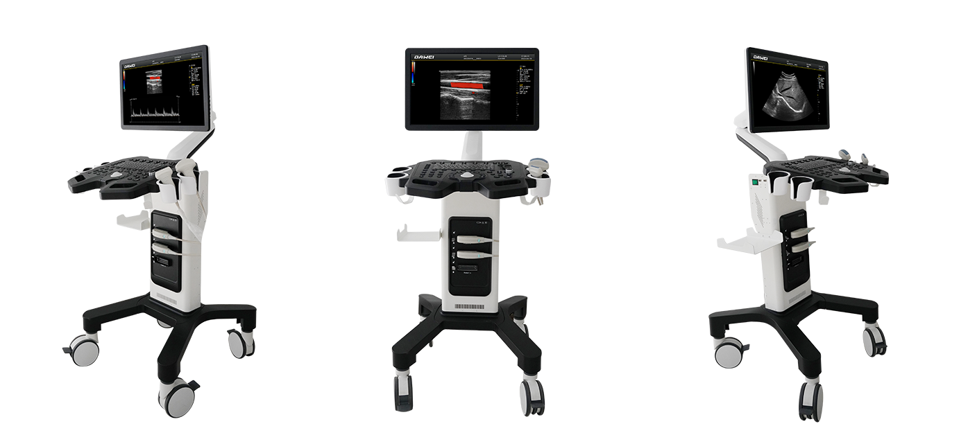
Appearance Display
Appearance
Flexible and fast
Technical Features
Stable, Durable, Stylish and Simple
21.5-Inch High-Resolution Medical Display
Flexible and Convenient Adjustable Arm
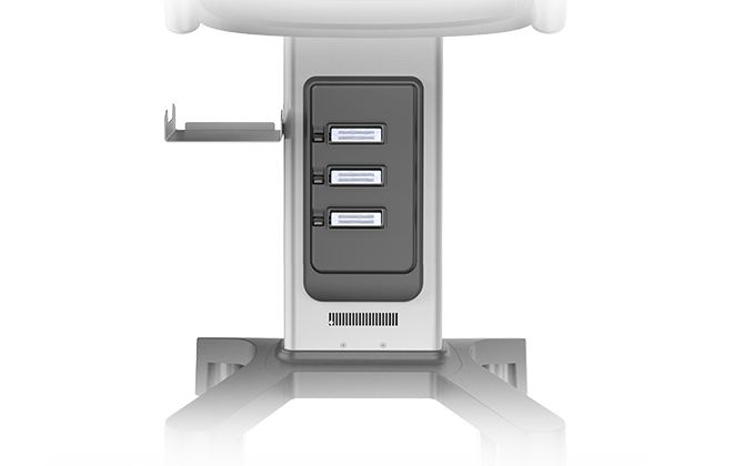
Integrated Backlit Silicone Keyboard
Fully activated three-probe interface
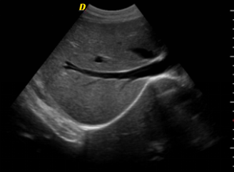
Spatial Compound Imaging Technology
Ultrasonic spatial compound imaging can improve contrast resolution, fine resolution and spatial resolution Enhance tissue and lesion interface echo continuity, reduce various artifacts (specular reflection, speckle, scattering, attenuation, poor contrast)
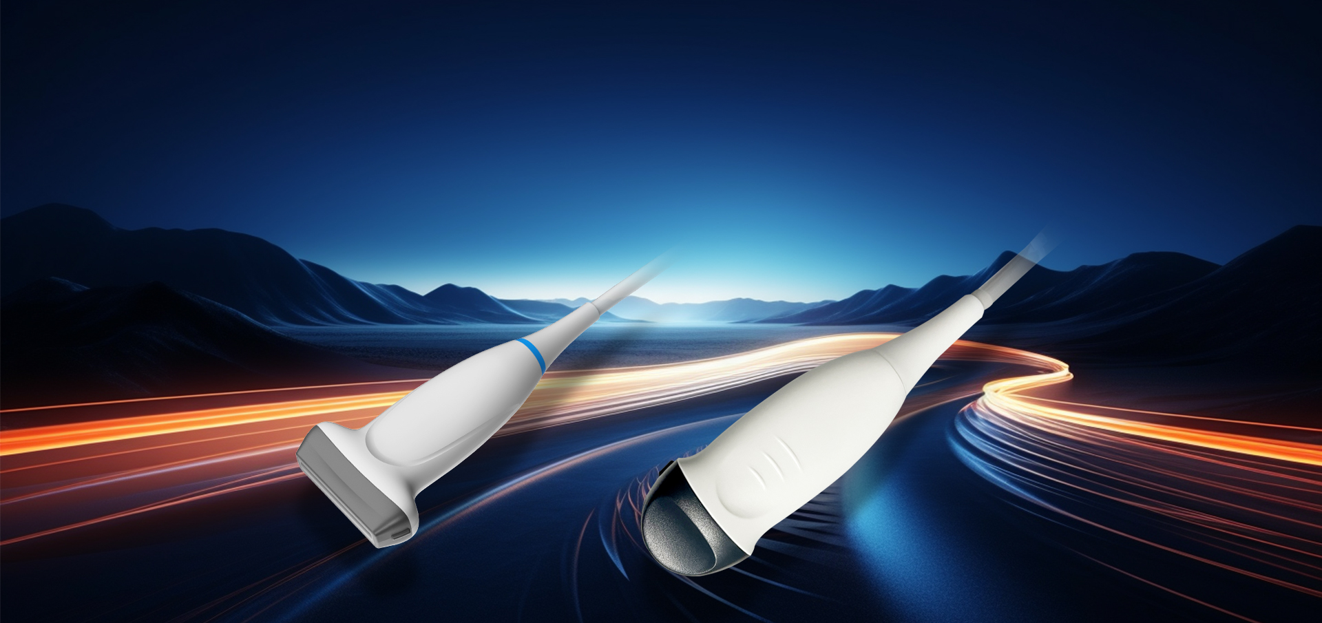
Probe
Advanced Probe Applications
The probe is made from composite materials, offering superior penetration and higher resolution. The images are consistently uniform without any loss of detail, providing more than 60% improvement in performance compared to traditional piezoelectric ceramic materials。
Sub-cutting allows for complete control of the entire process of chip vibration, reducing sidelobe artifacts, enhancing fine tissue resolution, and achieving sharper boundaries between adjacent strong echo reflectors. This fully showcases the high-resolution images brought by the high-density probe, perfectly presenting image details, and increasing clinical diagnostic accuracy.
Lossless Signal Transmission and Reception
High Precision A/D Conversion
Spatiotemporal Wave Velocity Reconstruction
Image Post-Processing
Unique pure wave probe technology
Bring high resolution images
Featuring a dedicated high-density probe, employing a completely new array design technology and an unique pure wave probe technology, undergoes secondary cutting of individual transducers, allowing for full control over the transducer's vibration throughout the process. This reduces sidelobe artifacts, enhances the resolution of fine tissue details, and sharpens the boundaries between adjacent strong echo reflectors. It fully showcases the high-resolution images brought by the high-density probe, perfectly presenting image details, and increasing the accuracy of clinical diagnosis.