Veterinary Color Doppler Ultrasound Diagnostic System
Started with Original Intent, achieved with craftsmanship
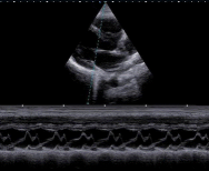
High-definition Clinical Images
Rich Configuration Expansion
Comprehensive Function Applications
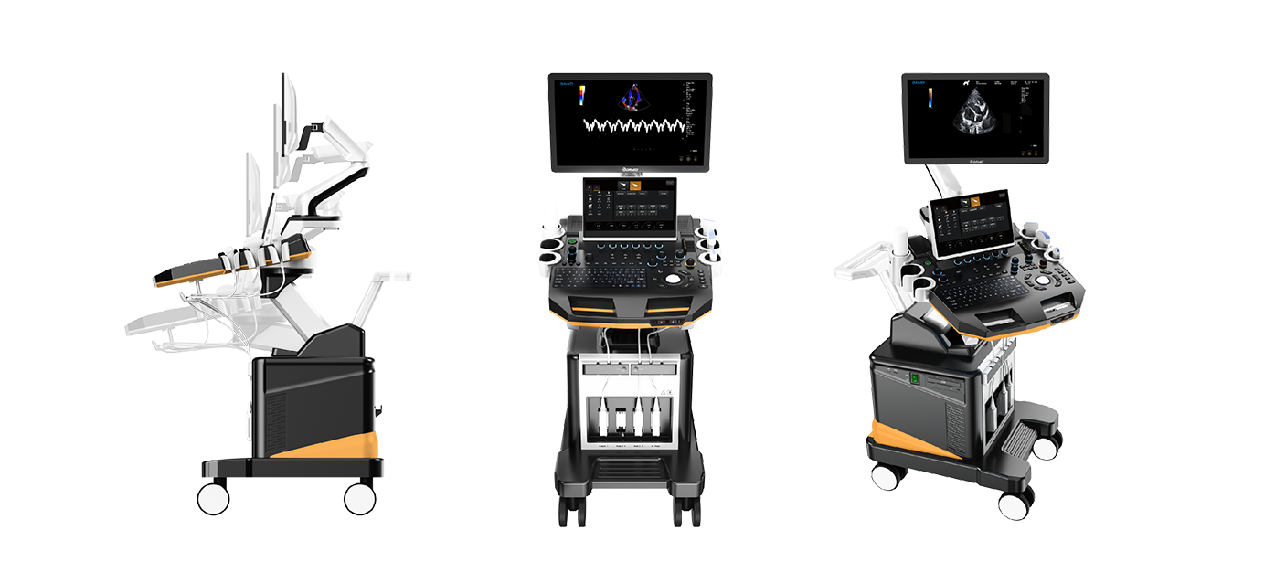
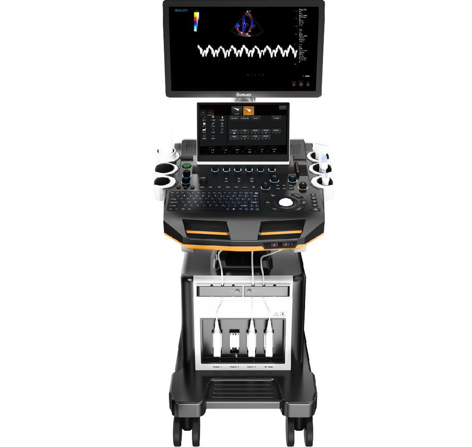
Appearance Display
Appearance
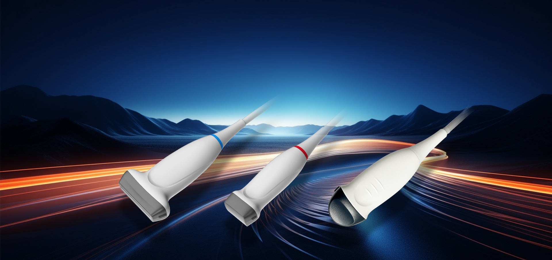
Probes
Technical Features
Started with Original Intent, achieved with craftsmanship
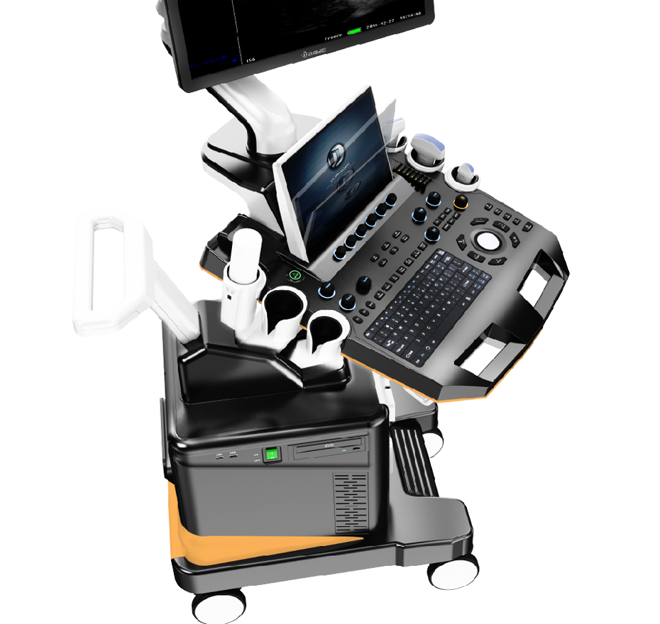
Panel "bearing" Design
Touch Screen 0-45° Angle Adjustment
Height-adjustable Button
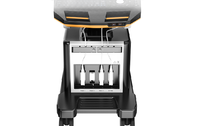
Four active probe interface
Meet the needs of a variety of scenarios, multi-functional general practice clinical application
Anatomic M-mode Imaging
Traditional M-mode has only one M-mode sampling line, which has limitations for examining tissue in motion, especially in difficult patients。 Anatomic M mode makes up for the shortcomings of traditional M mode in examining difficult patients by providing multiple M sampling lines, allowing you to perform more effective motion analysis of M mode at different angles and positions
Probe
Advanced Probe Applications
The probe is made from composite materials, offering superior penetration and higher resolution. The images are consistently uniform without any loss of detail, providing more than 60% improvement in performance compared to traditional piezoelectric ceramic materials.
Sub-cutting allows for complete control of the entire process of chip vibration, reducing sidelobe artifacts, enhancing fine tissue resolution, and achieving sharper boundaries between adjacent strong echo reflectors. This fully showcases the high-resolution images brought by the high-density probe, perfectly presenting image details, and increasing clinical diagnostic accuracy.
Lossless Signal Transmission and Reception
High Precision A/D Conversion
Spatiotemporal Wave Velocity Reconstruction
Image Post-Processing
Unique pure wave probe technology
Bring high resolution images
Featuring a dedicated high-density probe, employing a completely new array design technology and an unique pure wave probe technology, undergoes secondary cutting of individual transducers, allowing for full control over the transducer's vibration throughout the process. This reduces sidelobe artifacts, enhances the resolution of fine tissue details, and sharpens the boundaries between adjacent strong echo reflectors. It fully showcases the high-resolution images brought by the high-density probe, perfectly presenting image details, and increasing the accuracy of clinical diagnosis.